In this lesson, we’ll take a look at the generation of a brain wave, tying in concepts from the basics of cellular neuroscience and the electrophysiology of EEG.
The Firing Neuron
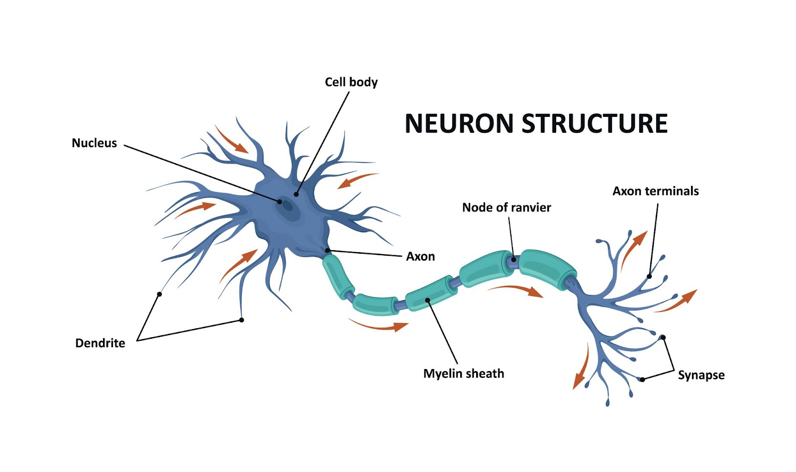
The basic functional unit of the nervous system is the nerve cell, or neuron. The brain contains on the order of 100 billion neurons, with each neuron on average connecting with approximately 10,000 other neurons. Neurons connect through synapses to form neural circuits, which mediate functions spanning from simple reflexes to perception, learning, and behavior. Neural circuits connect with each other to form large-scale brain networks, whose emergent properties function to produce complex cognition and behaviors, and when compromised, disease states.
Neural communication occurs through a complex interaction of chemical and electrical signals.
Nerve impulses, otherwise known as action potentials, are the all-or-none electrical events that originate from thousands of inputs to a neuron at its branch-like extensions called dendrites. Once a threshold has been reached, this information is sent from the cell body down the axon to the terminal buttons. The axon may be insulated with a lipid-rich material known as myelin for faster transmission, or unmyelinated. Myelinated axons are the source of the term white matter. Myelination is a process that occurs with maturation in early brain development.
At the molecular level, the transmission of a neural impulse is dependent on the movement of ions which increase (or depolarize) the membrane of the neuron. The signal travels down the axon in a bucket brigade manner as each segment of the membrane depolarizes the neighboring membrane. Once the signal reaches the terminal buttons, it stimulates the release of neurotransmitters into the synapse, the space that separates the presynaptic neuron from the postsynaptic neuron. This excites (or inhibits, depending on the neurotransmitter) dendrites of the postsynaptic neuron. In the case of an excitatory postsynaptic potential, if enough simultaneous signals are integrated at this neuron, another action potential will be generated and the process will continue. Nearly 80% of neurons are excitatory, but the interplay between inhibition and excitation plays a key role in the functioning of circuits, and imbalances in this ratio underlie neurological disorders such as epilepsy.
How do Action Potentials Create a Brain Wave?
In the process of creating an action potential, the movement of ions during an impulse creates a temporary separation of charge across the neuronal membrane. This separation of charge results in the neuron acting like an electrical dipole, which you can think of as a small battery with a positive and negative pole. This means there is an electric current fluctuating not only in the neuron but also in the surrounding extracellular space. Cerebral tissue/fluid acts like a conducting medium of these currents, in a process known as volume conduction. This is how potentials reach the scalp.
EEG or electroencephalogram, (literally meaning electric brain graph) provides a non-invasive way to view brain function by mapping the electrical activity of large numbers of neurons using electrodes placed on the scalp. Instead of sensing the electrical activity associated with action potentials which happen on the order of one millisecond, EEG senses the postsynaptic potentials which can last for up to several tens of milliseconds (Kirschstein and Kohling 2009).
EEG signals that you can measure come from when large numbers of pyramidal neurons sum their excitatory and inhibitory potentials to a “consensus” potential, which is a total sum of many small potentials from these neurons. Recall excitatory postsynaptic potentials (EPSPs) make the postsynaptic neuron more likely to fire an action potential, while inhibitory postsynaptic potentials (IPSPs) make it less likely.
Cortical neurons are arranged in a columnar fashion that is perpendicular to the surface of the cortex. As a consequence, depolarizations at the surface and deep levels of the columns cause different EEG signals. To an EEG, surface IPSPs and deep EPSPs look the same and produce positive (downward) deflections in the EEG, and surface EPSPs and deep IPSPs look the same and produce negative (upward) deflections in the EEG.
The postsynaptic potentials can sum spatially as well as temporally in a process known as synchronization. If multiple neurons in a region receive synchronized inputs, the postsynaptic potential may sum, leading to a synchronized response. Similarly, if neurons receive repeated inputs at a particular frequency, the postsynaptic potentials may overlap and sum, leading to synchronized activity.
For more information, we recommend checking out this article from LearningEEG on the basics of EEG electrophysiology. In the following five lessons, we will explore each brain wave in detail.