In this lesson, we’ll take a broad tour of the human brain, the most complicated and mysterious piece of matter in the known universe. Let’s begin by examining the general organization of the human brain.
The brain can, most broadly, be divided into the brain stem, cerebellum, and cerebrum. The brain stem links the brain to the spinal cord and controls primitive regulatory functions such as breathing, heart rate, blood pressure, and sleep. Behind the cerebrum lies the cerebellum (derived from Latin for little brain), which plays a key role in movement and connects extensively with the cerebrum and spinal cord.
The cerebrum is the largest, frontal-part of the brain that is split into two cerebral hemispheres, separated by the sagittal fissure which divides the two hemispheres. This bilateral symmetry means nearly every structure in the brain comes in pairs. The outer shell of the cerebrum is known as the cerebral cortex. You can visualize the cerebral cortex like a crumpled sheet composed of gray matter that has an area roughly the size of a dinner napkin.

The cerebral cortex is organized into six stacked layers and divided into four lobes. Under the frontal bone of the forehead lies the frontal lobe, which accounts for nearly 30% of the neural real estate in the brain. It is the primary processing center for attention, memories, and high level processing like executive functioning. The central sulcus is a deep indentation that divides the frontal and parietal lobe, found at the posterior border of the frontal lobe. The parietal lobe processes sensory inputs from the skin as well as spatial navigation and bodily orientation in space (proprioception). Posterior to the parietal lobe at the back of the brain is the occipital lobe, which handles visuospatial processing. Beneath the temples lies the temporal lobe, which primarily processes hearing, memories, and emotions.
Let’s map this knowledge over to the domain of EEG practice. The International 10-20 system assigns each region of the cerebral cortex by a letter and number. Each point below designates an area of the scalp an electrode may be placed:
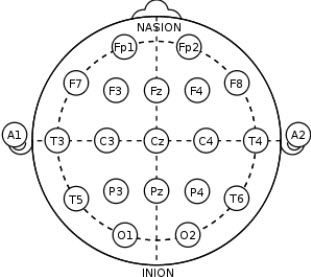
- F, frontal lobes
- Fp, frontal poles
- P, parietal lobes
- T, temporal lobes
- Tp, temporal poles
- O, occipital lobes
- C, central/sensorimotor cortex
- Z, the midline dividing the hemispheres
Each letter before the number refers to a cortical lobe. Odd numbers refer to the left side of the brain, while even numbers refer to the right side of the brain.
Now, in order to describe the locations and relationships between these structures, it’s useful to unpack the terminology of anatomical directions.

- Posterior: toward the back of the head
- Anterior: toward the front of the head
- Ventral: toward the bottom of the head
- Dorsal: toward the top of the head
- Medial: midline of the brain
- Superior: nearer the top (dorsal)
- Inferior: nearer the bottom (ventral)
- Vertex: central position, Cz, Pz, Fz
- Lateral: to the left or right of midline
Other useful terminology
Contralateral refers to locations on opposite sides. For instance, the left ear is contralateral to the right ear.
Ipsilateral means a given structure or site is limited to just one part of the head. Site F4 is ipsilateral to C4.
Homologous sites are 10-20 sites that are complementary across hemispheres. For instance, F3 and F4 are homologous, as is C3 and C4.
Left vs Right Hemisphere
The split-brain theory dates back to the work of Roger Sperry in the 1960s and 1970s, who won a Nobel Prize for his work. Sperry had a patient with intractable epilepsy that was treated, as a last resort, by severing the corpus callosum, the main superhighway of the brain that allows cross-talk between the hemispheres. After the operation, what Sperry found was remarkable: when the hemispheres are not connected to each other, they function independently as if the patient had two brains in one body. Naturally, this had some interesting implications.
One of the main consequences of this work was that Sperry found that each hemisphere is specialized for different functions. For instance, the production of language appears to be a left-hemispheric function. If a split-brain patient is shown an image or object on the left visual field, they can’t articulate what they’ve seen but they can identify the object with solely their left hand. This is because the right hemisphere processes the left visual field, but this hemisphere doesn’t process language (but does of course process muscle functions on the left side of the body).
Also, as opposed to the left hemisphere’s focus on small details and quantitative logic, the right hemisphere appears to be more involved in seeing the big picture, making associations between information and holding a gestalt view of objects and the environment. As such, the right hemisphere is intimately connected with intuition and creativity. That being said, the idea of right or left-brain dominated thinking is simplistic and doesn’t capture the reality that both sides of the brain always work together as a system.